An intraoral camera is a compact, pen-sized imaging device designed to capture detailed photographs and video of the teeth, gums, and other soft tissues inside the mouth. Unlike an ordinary camera, it is built specifically for intraoral use: it produces full-color, high-resolution images in real time and transmits them directly to a computer monitor so both clinician and patient can see exactly what the camera shows. The clarity of these images makes subtle changes and small lesions far easier to spot than with visual inspection alone.
Modern intraoral cameras incorporate bright, focused lighting and carefully engineered optics to reduce glare and reveal texture, color, and surface detail. Because the camera can be positioned within millimeters of a tooth surface, it can expose fine cracks, early decay, erosion, and marginal breakdown around restorations that might otherwise be missed. Images are typically captured as stills or short video clips and can be reviewed frame-by-frame to support diagnosis.
Beyond the hardware itself, the value of an intraoral camera rests in its ability to create a clear, objective record. Digital images become part of the patient’s chart, allowing clinicians to track changes over time, compare anatomy from visit to visit, and share visuals with other specialists when collaborative care is needed. This combination of precision imaging and easy documentation strengthens clinical decision-making at every stage of treatment.
One of the most immediate benefits of intraoral imaging is the way it transforms conversations between clinician and patient. Instead of relying on verbal descriptions or a clinician’s interpretation, patients see high-quality images of their own mouths, making observations concrete and easier to understand. This visual approach helps patients grasp the nature and location of concerns, why certain treatments are recommended, and how oral health can change over time.
Seeing is often more persuasive than being told, especially when it comes to subtle issues like hairline cracks, stain versus decay differentiation, or the exact edge of a filling. When patients can view and discuss the same images with the dental team, questions become more focused and informed consent is better supported. That shared view also encourages collaborative decision-making and helps patients feel more engaged in their care.
Clinicians can annotate images, draw attention to specific areas, and compare photos taken at different visits, which reinforces learning and follow-through. For families and caregivers, these visuals simplify explanations and make it easier to monitor pediatric or geriatric patients’ progress. The immediate, visual feedback is a powerful educational tool that improves understanding without oversimplifying important clinical details.
Intraoral cameras enhance early detection by revealing surface details that are difficult to see with the naked eye. Early caries, microfractures, failing margins around crowns or fillings, and initial signs of wear or erosion are all more easily identified when magnified and well-illuminated. Catching these issues sooner often translates into simpler, more conservative treatment and better long-term outcomes for the tooth.
Because images can be saved and compared over time, clinicians can track the progression—or stability—of suspicious findings. This temporal information helps distinguish between a longstanding, stable feature and a changing lesion that requires intervention. For restorative planning, high-quality images also assist in mapping the exact location and extent of defects, supporting more precise and predictable treatment results.
In practices that coordinate with specialists, intraoral photographs provide a standardized, shareable format for consultation. When an endodontist, periodontist, or lab technician needs to review an issue, having accurate intraoral images streamlines communication and reduces ambiguity. That clarity supports better interdisciplinary planning and often shortens the time between diagnosis and effective treatment.
An intraoral camera is not a standalone treatment but a complementary diagnostic and communication tool that integrates with modern dental workflows. During routine exams, it becomes part of a multi-faceted assessment along with visual inspection, tactile exploration, and, when appropriate, radiographs or CBCT imaging. Together, these tools provide a fuller picture of oral health and help the team select the right next steps.
Images captured by the intraoral camera are typically archived in the patient’s digital record, which helps the care team maintain continuity across visits. Whether monitoring a restoration, documenting the need for referral, or educating a patient about oral hygiene priorities, the images enhance clinical records and make future comparisons straightforward. This integration supports both preventive and restorative strategies by making clinical history more transparent and accessible.
For patients considering elective or cosmetic procedures, intraoral photography also aids in treatment planning by offering a clear baseline. It can be used to illustrate expected changes and to document outcomes once treatment is complete. Throughout the process, the camera’s images help ensure that recommendations are evidence-based, personalized, and explainable in plain terms.
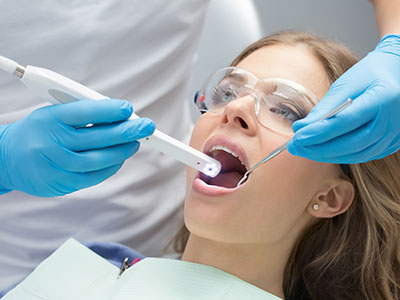
Undergoing an intraoral camera exam is quick, comfortable, and noninvasive. The device is smaller than many common dental instruments and is guided gently into the mouth while the clinician focuses on specific teeth or soft tissue areas. Bright but safe LED lighting illuminates the target, and images appear on a nearby monitor in real time so you can follow along as they are captured.
The procedure typically adds only a few minutes to a standard dental visit. Because images are digital, they can be adjusted for contrast and magnification on the screen to highlight areas of interest without needing repeat captures. If the clinician identifies something that warrants closer attention, additional photos may be taken from different angles or at higher magnification to ensure a comprehensive record.
After the images are taken, your dentist will review them with you, explaining what each image shows and how it factors into your overall care plan. Any findings that require follow-up—whether monitoring, restorative treatment, or referral—will be explained in clear terms. The visual documentation also becomes part of your chart, so it is available for future reference and for coordination with other dental or medical providers when necessary.
At Ballston Dental Center, our team uses intraoral imaging to make exams more transparent and to support informed, evidence-based care. If you’d like to learn more about how intraoral cameras are used during appointments or how images may be included in your treatment records, please contact us for more information.


A intraoral camera is a compact, pen-sized imaging device designed to capture high-resolution color photographs and short video of the teeth, gums and other soft tissues inside the mouth. The device transmits images in real time to a nearby monitor so clinicians and patients can view the same visuals together. These images can be captured as stills or clips and reviewed frame-by-frame to support diagnosis.
Modern intraoral cameras use focused LED lighting and precision optics to reduce glare and reveal fine surface detail such as texture, color variation and subtle defects. Because the camera can be positioned within millimeters of a tooth, it can expose hairline cracks, early decay and marginal breakdown around restorations that are difficult to see with visual inspection alone. The combination of magnification and illumination makes small changes far easier to detect and document.
Intraoral images provide objective, high-detail documentation that supports more accurate diagnosis and conservative treatment planning. By revealing early caries, microfractures and worn margins, the camera helps clinicians choose less invasive interventions when appropriate and target treatment to the exact location of concern. Saved images also enable clinicians to compare findings over time and verify whether a condition is stable or progressing.
The visual record is valuable when mapping restorative work because photographs clarify the extent and exact position of defects for both the dentist and the laboratory. Annotating images and pairing them with clinical notes improves communication within the dental team and with outside specialists. That precision reduces ambiguity and promotes more predictable treatment outcomes.
An intraoral camera exam is noninvasive and generally well tolerated by patients of all ages. The device is smaller than many dental instruments and is guided gently into the mouth while focused LED lighting provides illumination without discomfort. Because it captures visual images rather than using ionizing radiation, the camera is a complementary tool to radiographs rather than a replacement.
Typical intraoral imaging adds only a few minutes to a standard dental visit and does not require special preparation from the patient. Infection-control protocols and barrier covers are used for each exam to maintain hygiene and safety. The quick, painless nature of the procedure makes it a practical addition to routine exams and restorative evaluations.
Images taken with an intraoral camera are digitized and archived in the patient’s electronic dental record to create a clear visual history. These records allow clinicians to track changes over time, compare anatomy from visit to visit and reference prior findings during follow-up appointments. Digital files can also be exported or shared with outside providers when collaborative care or specialist consultation is needed.
At Ballston Dental Center, intraoral photographs are maintained as part of each patient’s chart to support continuity of care and evidence-based decision making. Image storage and sharing are handled securely and in accordance with applicable privacy standards so visuals are available for clinical review without compromising confidentiality. Having a visual record makes patient education and long-term monitoring more transparent and actionable.
Yes. By providing magnified, well-illuminated views of tooth surfaces, intraoral cameras make it easier to spot early enamel breakdown, staining patterns consistent with decay and tiny fractures that may not be visible on a cursory exam. Early detection often allows clinicians to recommend preventive or minimally invasive treatments that preserve more natural tooth structure. Photographs also help distinguish between surface stains and areas that require restorative care.
Because images can be saved and compared over time, the camera is particularly useful for monitoring suspicious areas rather than rushing into treatment. Clinicians can document whether a lesion is stable or showing change and use that temporal information to guide timely intervention. This approach supports conservative dentistry and better long-term outcomes for affected teeth.
An intraoral camera puts the same high-quality images in front of both clinician and patient, which transforms abstract descriptions into concrete visuals. When patients can see the exact area of concern, explanations about diagnosis and recommended treatments become easier to understand and questions become more focused. This shared visual context strengthens informed consent and helps patients feel more engaged in care decisions.
Clinicians can annotate images, highlight margins or fractures and compare current photos to prior visits, which reinforces learning and follow-through. For families and caregivers, the ability to view and discuss images simplifies explanations about pediatric or geriatric oral health needs. Overall, visual feedback improves clarity without oversimplifying important clinical detail.
Intraoral cameras capture surface color, texture and fine topographic detail that radiographs and CBCT scans cannot show, while X-rays and CBCT provide information about internal structures such as bone levels, root anatomy and interproximal decay. Because each modality reveals different aspects of oral health, using them together creates a fuller diagnostic picture. Surface images clarify what the clinician sees during an exam and can indicate when additional radiographic evaluation is warranted.
For restorative planning and interdisciplinary cases, combining intraoral photographs with radiographic data improves precision and communication. Photographs show the clinical appearance and margin details while radiographs confirm underlying structure and pathology. This complementary approach reduces uncertainty and supports more informed clinical decision making.
The exam is quick, comfortable and noninvasive; the clinician will guide the pen-sized camera into the mouth and focus on teeth or soft tissue areas of interest while images appear on a monitor in real time. Bright but safe LED lighting illuminates the target and allows the clinician to capture multiple angles and close-up views as needed. Because images are digital, they can be adjusted for contrast and magnification on screen to highlight areas of concern without repeat captures.
The process typically adds only a few minutes to a routine appointment, after which the dentist will review the images with you and explain their significance for your care plan. If additional photos are needed to document a restoration or to support a referral, the clinician will take them and archive them in your record. Having the images available makes follow-up conversations and comparisons at future visits straightforward.
Intraoral photographs provide a standardized, shareable visual format that specialists and laboratory technicians can review to understand clinical appearance and precise locations of concern. When referring to an endodontist, periodontist or oral surgeon, sending clear images alongside clinical notes reduces ambiguity and speeds up collaborative planning. For laboratory work, photographs help technicians match shades, assess margins and fabricate restorations that fit the documented clinical situation.
Because images are archived in the patient record, they become a reliable reference during interdisciplinary consultations and reduce the need for repeated chairside explanations. Sharing visuals helps ensure all members of the care team are aligned on the problem and intended outcome. That alignment supports efficient communication and more predictable treatment coordination.
Ballston Dental Center incorporates intraoral imaging to increase transparency, improve diagnostic accuracy and support evidence-based treatment planning during routine exams and restorative visits. The technology enhances patient education by showing real-time visuals and allows clinicians to document and monitor oral health changes over time. Using intraoral cameras as part of a multi-modal diagnostic approach supports early detection and more conservative care when appropriate.
Images captured during visits are added to the patient’s digital record to promote continuity of care and to simplify coordination with specialists when needed. This visual documentation helps the dental team communicate findings clearly and make recommendations that are explainable in plain terms. The result is a more informed, collaborative approach to maintaining and restoring oral health.
We are here to help you take the next step toward a healthier smile.
Whether you have questions about our services, insurance, or scheduling your first visit, our team is ready to assist you.
We welcome new and returning patients and are happy to help you find a time that fits your schedule. Reach out to our office today to schedule an appointment or learn more about how we can support your oral health. We look forward to welcoming you to our practice.