At the office of Ballston Dental Center, we invest in diagnostic technologies that help clinicians see more, plan more precisely, and deliver care with greater confidence. Cone-beam computed tomography (CBCT) is one of those tools: a three-dimensional imaging modality that reveals anatomy in a way conventional two-dimensional X-rays cannot. When used thoughtfully, CBCT supports clearer diagnoses and more predictable treatment planning across a wide range of dental specialties.
CBCT is not a replacement for routine X-rays, but a complementary technology reserved for cases where extra information makes a meaningful difference in outcomes. Our team follows evidence-based guidelines to determine when a scan is appropriate, and we integrate each CBCT study into a broader diagnostic process designed to prioritize safety, comfort, and clinical clarity.
Traditional bitewing and panoramic X-rays provide valuable snapshots of tooth structure and surrounding bone, but they compress complex anatomy into flat images. CBCT produces volumetric datasets that allow clinicians to view teeth, bone, nerves, and sinuses from multiple angles and in cross-section. This perspective makes it easier to detect subtle patterns of disease, assess spatial relationships, and identify anatomical variations that could affect treatment.
Because CBCT captures depth and orientation, it improves the ability to locate root canals, detect vertical fractures, and evaluate the full extent of lesions that might be obscured on 2-D films. It also clarifies the relationship between tooth roots and adjacent structures such as the mandibular nerve or the maxillary sinus—information that is essential when evaluating risk and planning interventions.
Beyond immediate dental structures, CBCT can reveal bone volume and quality, the presence of accessory canals, and other conditions that influence long-term prognosis. Having this level of detail up front helps clinicians avoid surprises during treatment and communicate expectations more accurately with patients.
Surgical precision depends on accurate visualization. For dental implant planning, CBCT allows for measurement of bone height, width, and density in three dimensions so clinicians can determine optimal implant size and angulation. Knowing the exact location of nerves and sinus cavities reduces the risk of complications and helps guide minimally invasive approaches when appropriate.
CBCT datasets are often used with planning software to simulate implant placement and, when indicated, fabricate surgical guides that transfer the virtual plan to the mouth. This digital workflow enhances reproducibility and helps ensure implants are positioned for long-term function and esthetics while preserving vital anatomy.
For oral surgery beyond implants—such as impacted tooth removal or cyst evaluation—CBCT gives surgeons a clearer map of the surgical field. That clarity allows for more conservative bone removal, targeted incisions, and shorter operating times, all of which contribute to improved patient recovery when combined with sound clinical judgment.
Endodontists rely on CBCT to examine complex root anatomies, identify missed canals, and locate regions of resorption or persistent infection that are difficult to appreciate on conventional films. When retreatment is being considered or anatomy is ambiguous, a CBCT scan can be decisive in choosing the safest, most effective course of care.
Orthodontic and restorative teams also benefit from 3-D imaging when evaluating impacted teeth, planning extractions, or assessing skeletal relationships. CBCT assists in visualizing tooth inclinations, root proximity, and alveolar boundaries, enabling multidisciplinary planning that balances tooth movement with periodontal health.
In airway and sleep-related assessments, CBCT can provide anatomic information about the nasal passages, pharyngeal airway volume, and surrounding structures. While CBCT is only one part of a comprehensive airway evaluation, the images can help clinicians identify anatomical contributors to breathing issues and work with medical colleagues to develop coordinated care plans.
Radiation exposure is an important consideration with any imaging study. CBCT units used in dental practices are designed to deliver substantially lower doses than conventional medical CT scans because they focus on a limited field of view and use dental-optimized protocols. We select the smallest field and lowest exposure consistent with the diagnostic task to reduce dose while preserving image quality.
CBCT scans are also fast. Most dental CBCT acquisitions take only a few seconds, which reduces motion artifact and improves patient comfort. Because the procedure is noninvasive and completed with the patient seated or standing in an open unit, many patients find it easier to tolerate than enclosed medical imaging devices.
Our team follows strict quality control and positioning procedures to ensure each scan is clinically useful. We also review indications for scanning carefully, reserving CBCT for situations where the additional detail will influence treatment decisions or outcomes.
Capturing a CBCT scan is only the first step. The real value comes from how images are interpreted and incorporated into the treatment plan. Our clinicians review CBCT data using specialized software that enables measurements, sectional views, and three-dimensional reconstructions. This allows for more precise diagnoses and helps the team anticipate challenges before treatment begins.
CBCT findings are discussed with patients in plain language, often with visual aids from the scan itself. Seeing a cross-sectional view or a 3-D model can make complex conditions easier to understand and supports shared decision-making. When multiple specialists are involved, the CBCT dataset serves as a common reference that streamlines communication and collaborative planning.
We also archive CBCT studies securely so they remain available for future comparison or referral. Having a baseline three-dimensional record can be invaluable for monitoring healing, assessing progression, or coordinating follow-up care when needed.
In summary, CBCT adds a dimensional advantage to dental diagnosis and treatment planning—helping clinicians visualize anatomy more completely, reduce uncertainty, and design care with greater precision. If you have questions about whether CBCT is appropriate for your situation or what to expect during a scan, please contact Ballston Dental Center to learn more.


Cone-beam computed tomography (CBCT) is a three-dimensional imaging modality that captures volumetric data of the teeth, jaws, and surrounding structures. Unlike traditional two-dimensional bitewing or panoramic X-rays, CBCT provides cross-sectional views and 3-D reconstructions that reveal depth, orientation, and spatial relationships. This added perspective makes it easier to detect root fractures, assess bone volume, and visualize the proximity of nerves and sinuses.
Because CBCT images are three dimensional, clinicians can measure distances and angles with greater accuracy than on flat films. The technology complements rather than replaces routine radiographs and is typically used when extra detail will influence diagnosis or treatment planning. Proper interpretation requires specialized software and training to convert the volumetric dataset into clinically useful views.
CBCT is recommended when two-dimensional imaging does not provide sufficient information for safe, predictable treatment. Common indications include implant planning, evaluation of impacted or atypically positioned teeth, assessment of complex root anatomy, and surgical mapping for extractions or cyst removal. It is also used selectively for airway assessment and certain diagnostic dilemmas where three-dimensional detail changes clinical decisions.
Clinicians follow evidence-based guidelines to weigh benefits against radiation exposure and to choose the smallest field of view needed for the task. CBCT is not a routine substitute for bitewings or panoramic films but a targeted tool reserved for situations in which the additional information will affect outcomes. Shared decision-making with the patient helps determine whether a scan is appropriate for a given case.
CBCT allows clinicians to evaluate bone height, width, and density in three dimensions, enabling precise selection of implant size, angulation, and position. The images show the exact relationship of tooth roots to adjacent anatomical structures such as the mandibular nerve and the maxillary sinus, which reduces the risk of intraoperative complications. Many practices use CBCT datasets with planning software to simulate placement and design surgical guides that translate the virtual plan into accurate clinical execution.
For other oral surgery procedures, CBCT provides a detailed map of the surgical field that supports more conservative bone removal and targeted incisions. This clarity can shorten operative time and help anticipate challenges like aberrant anatomy or accessory canals. When integrated into a broader treatment plan, CBCT contributes to more predictable healing and long-term function.
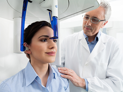
A dental CBCT scan is typically quick and noninvasive, with most acquisitions completed in a few seconds while the patient is seated or standing. The patient is positioned in the unit and instructed to remain still while the machine rotates around the head, capturing the volumetric dataset. Because the device is open and the exposure time is short, many patients find the experience more comfortable than enclosed medical CT scanners.
After the scan, images are processed and reviewed by the clinician using specialized software that produces sectional views and 3-D reconstructions. The clinician will explain relevant findings in plain language and use images to illustrate anatomy or treatment options when appropriate. Scans are also archived for future comparison or for referral if multidisciplinary care is needed.
Radiation dose for dental CBCT is substantially lower than that of conventional medical CT scans because dental units use a focused field of view and dental-optimized protocols. Clinicians minimize exposure by selecting the smallest field and lowest effective settings necessary for the diagnostic task. Modern CBCT units and adherence to ALARA (as low as reasonably achievable) principles help balance diagnostic benefit with radiation safety.
Certain populations, such as pregnant patients, require special consideration and discussion of risks and benefits before imaging. Your clinician will review clinical need and, when appropriate, suggest alternative diagnostic strategies or delay nonurgent imaging. Proper indication, technique, and shielding when indicated all contribute to maintaining patient safety.
Endodontists use CBCT to visualize complex root canal anatomies, locate additional or missed canals, and detect fractures or areas of resorption that are not apparent on conventional films. Three-dimensional views can reveal the true extent of periapical lesions and help distinguish between anatomical variants and disease. This information often changes treatment approach, for example by indicating the need for retreatment, surgical intervention, or conservative monitoring.
When anatomy is ambiguous, CBCT supports case planning by showing canal curvature, root length, and the relationship of roots to nearby structures. The images improve diagnostic confidence and help clinicians counsel patients about prognosis and expected steps in treatment. All CBCT findings are interpreted in the context of clinical examination and patient symptoms.
In orthodontics and restorative dentistry, CBCT provides a comprehensive view of skeletal relationships, tooth inclinations, and root positions that can affect treatment selection and sequencing. The technology is particularly useful when assessing impacted teeth, evaluating cortical bone for planned tooth movement, or determining whether restorative procedures might compromise periodontal support. By visualizing roots and alveolar boundaries, clinicians can plan treatments that balance esthetics, function, and long-term tissue health.
Multidisciplinary teams often use CBCT datasets as a common reference to coordinate care between orthodontists, oral surgeons, and restorative dentists. The three-dimensional information supports more accurate risk assessment and can inform decisions such as staged extractions, orthognathic planning, or the timing of implant placement relative to tooth movement. As with other uses, the field of view is tailored to the clinical question to limit exposure.
CBCT can provide useful anatomic information about the nasal passages, pharyngeal airway volume, and surrounding structures that may contribute to breathing issues. The images help identify structural contributors such as enlarged adenoids, nasal obstruction, or reduced pharyngeal space that might be relevant to sleep-disordered breathing. However, CBCT is only one component of a comprehensive airway assessment and cannot replace clinical evaluation, sleep studies, or collaboration with medical specialists.
Clinicians use CBCT findings to inform referrals and to guide conservative or surgical interventions when anatomy is a clear contributing factor. Interpretation of airway metrics from CBCT should be done with caution because patient position and breathing state can affect measured volumes. Multidisciplinary coordination ensures that imaging findings are integrated with functional testing and medical history to form a complete care plan.
CBCT has limitations including lower soft-tissue contrast compared with medical CT and potential image artifacts from metal restorations or motion. It is not the ideal modality for evaluating soft-tissue pathology that requires high contrast resolution, and its diagnostic value depends on appropriate field selection and technique. Artifacts or improperly centered scans can reduce usefulness, so correct positioning and patient cooperation are important.
Additionally, CBCT should not be used indiscriminately; unnecessary imaging exposes patients to radiation without commensurate benefit. Clinicians weigh the expected impact of a scan on diagnosis and treatment before ordering it, and they may opt for conventional radiographs or alternative assessments when those are sufficient. Clear indication and careful interpretation maximize the clinical value of CBCT studies.
CBCT datasets are reviewed by the treating clinician using specialized viewing software that allows sectional analysis, measurements, and three-dimensional reconstructions. In complex or ambiguous cases, clinicians may consult or refer the study to a specialist such as an oral and maxillofacial radiologist, endodontist, or oral surgeon for a second opinion. Collaboration ensures that findings are interpreted accurately and that treatment plans reflect multidisciplinary expertise when appropriate.
Images from the scan are used to explain diagnoses and treatment options to patients with visual aids that make anatomy easier to understand. Scans are also securely archived for future comparison, which helps monitor healing and supports coordinated care over time. If you have questions about whether CBCT is appropriate for your situation, contact Ballston Dental Center to discuss indications and next steps.
We are here to help you take the next step toward a healthier smile.
Whether you have questions about our services, insurance, or scheduling your first visit, our team is ready to assist you.
We welcome new and returning patients and are happy to help you find a time that fits your schedule. Reach out to our office today to schedule an appointment or learn more about how we can support your oral health. We look forward to welcoming you to our practice.